What is the Dermatoscopy Method?
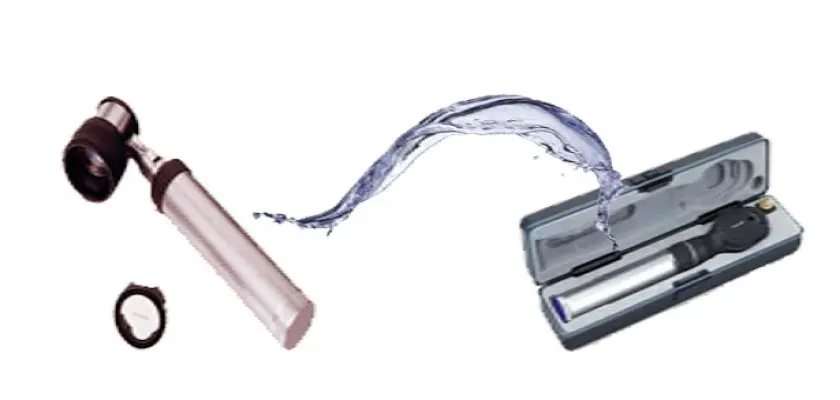
Dermatoscopy (Epiluminescence Microscopy) is a method used for examining and monitoring skin spots and moles (Melanocytic Nevus). It is also known as Dermoscopy. The dermatoscopy device magnifies the skin surface 10 to 100 times. This procedure, which allows for microscopic examination of the skin surface, enables the monitoring of changes in the structure of moles.
Early Diagnosis in Skin Cancer
In addition to structural changes in existing moles; the appearance of new moles on the body are symptoms of skin cancer. In this regard, moles on the bodies of individuals at risk of skin cancer should be recorded, and the aforementioned changes should be monitored. The dermatoscopy method allows for early diagnosis in skin cancer cases. Early diagnosis is life-saving, especially in melanoma type skin cancer. Melanoma type skin cancer, which grows rapidly and metastasizes without attracting attention, mostly appears as a mole. Again, early diagnosis is possible with a dermatoscopy device in skin cancers such as seborrheic keratosis, basal cell carcinoma, and squamous cell carcinoma, as well as dermatological diseases like scabies and alopecia areata. A sample is taken for biopsy from suspicious skin lesions diagnosed with the dermatoscopy method.